A live window beneath the skin
When a doctor looks at a face, they can see the skin, its shape and the way it moves. But much of the important anatomy is hidden underneath.
There are layers of skin, soft tissue and fat. There are muscles that create movement, bands of connective tissue that support the face, glands, and blood vessels carrying blood around the area. Every face contains these structures, but their exact position, depth and pathway can vary from one patient to another.
Ultrasound provides another source of information. It allows the Doctor to place a small probe on the skin and see a live, moving picture of some of the structures underneath.
It is a little like opening a window into the face without making a cut.
The very simple explanation: Ultrasound sends sound into the skin, listens for the echoes and turns those echoes into a picture.
What does the word “ultrasound” mean?
Humans can only hear sounds within a certain range. Ultrasound simply means sound with a pitch that is too high for our ears to hear.
The ultrasound probe sends these very high-frequency sound waves into the tissue. The sound waves travel until they meet something, such as skin, fat, muscle, a blood vessel or bone. Some of the sound then bounces back towards the probe as an echo.
A computer measures those echoes and quickly turns them into an image on the screen.
This happens many times every second, which is why the picture can move in real time. The doctor can move the probe, gently press on a structure, ask the patient to move a muscle or adjust the ultrasound settings and watch what happens immediately.
It is the same basic technology used to see a baby during pregnancy, although facial ultrasound uses a high-frequency probe designed to examine small, shallow structures close to the surface of the skin.
What happens during a facial ultrasound?
A facial ultrasound assessment is usually simple:
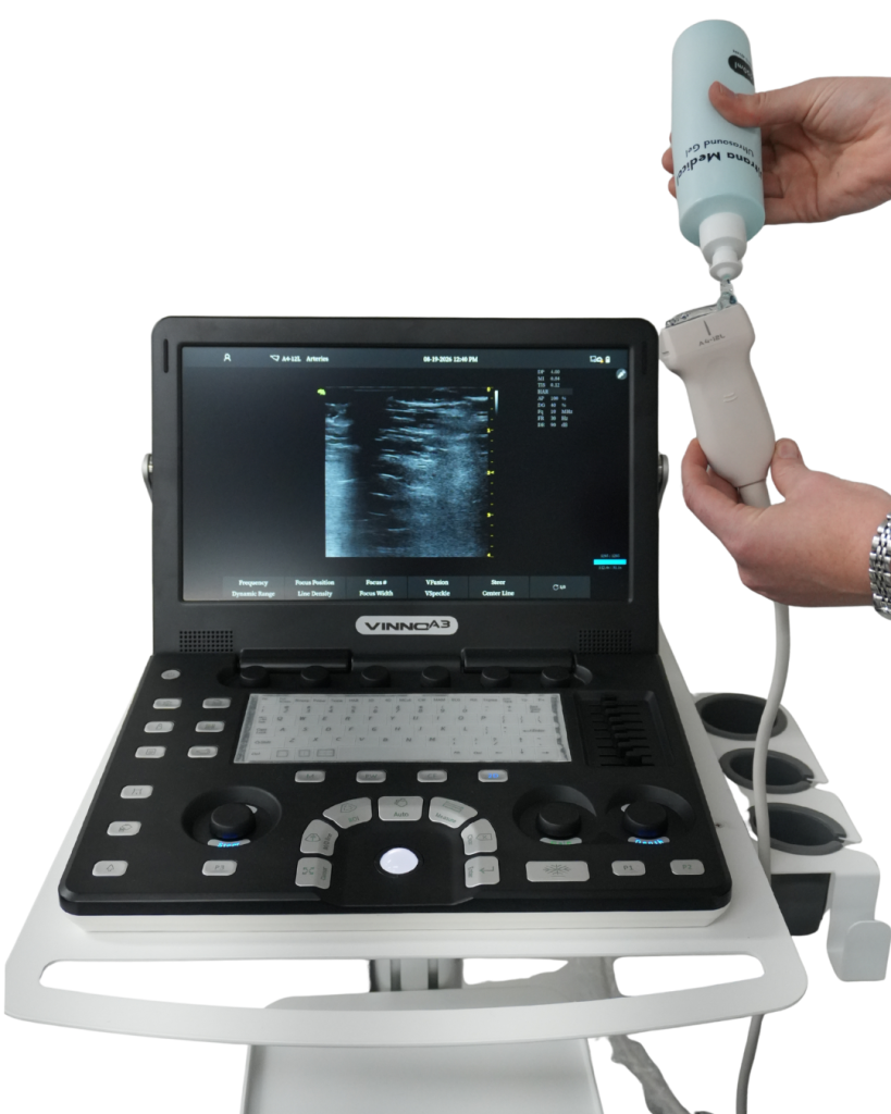
- A small amount of clear gel is placed on the skin.
- The ultrasound probe is rested gently against the face.
- The doctor moves the probe across the area being assessed.
- The anatomy appears as a moving black, white and grey image on the screen.
- Doppler settings may be used when information about blood flow is needed.
- Relevant findings can be considered alongside the patient’s history, examination and treatment plan.
The gel helps the sound travel between the probe and the skin. The scan itself does not involve an incision and diagnostic ultrasound does not use X-rays or ionising radiation.
Patients may feel the probe moving and some light pressure. The scan is generally not painful, although an area that is already swollen, inflamed or tender may feel sensitive when touched.
What can an Ultrasound see?
An ultrasound screen does not look like an ordinary photograph. It is more like a moving map made from different shades and patterns.
Depending on the area, the probe and the image quality, ultrasound may help visualise:
- The skin and dermal region
- Tissue immediately beneath the skin
- Fat compartments
- Fascia and other connective tissue
- Muscles
- Some glands
- Blood vessels and blood flow
- The bright surface of bone or cartilage
- Deposits from some previous treatments
- Fluid, swelling or other changes that may need assessment
Tendinous or fibrous structures may also be visible where they are present and relevant.
The face is curved and three-dimensional, while the ultrasound screen shows a thin slice at a time. The doctor builds an understanding by moving the probe through several positions, much like looking through the pages of a picture book one page at a time.
What do the colours mean?
On the usual greyscale image:
- Black areas often represent fluid or structures that return very few echoes.
- Grey areas represent different types of soft tissue.
- Bright white areas return strong echoes. Bone surfaces, fascia and other dense interfaces may appear bright.
When colour Doppler is switched on, colour can be placed over areas where the machine detects moving blood. The colours provide information about the direction and pattern of flow in relation to the probe. They are not simply labels for arteries and veins, so they still require skilled interpretation.
Why is ultrasound useful in facial medicine?
Doctors learn facial anatomy from textbooks, cadavers, clinical training and experience. That knowledge is essential, but a textbook shows a typical face. It cannot show exactly where a particular patient’s vessel travels today, what may remain from a previous treatment, or what lies beneath an area of swelling.
Ultrasound can add patient-specific, real-time information.
Think of it like driving with both a road map and a live view of the road. The map provides essential knowledge. The live view may show where things are in front of you right now.
Ultrasound may support:
- Assessment of anatomy in a planned treatment area
- Identification of some blood vessels and their pathway
- Understanding the depth and movement of facial muscles
- More individualised treatment planning
- Assessment of some previous injectable treatments
- Investigation of a lump, nodule, swelling or unexpected result
- More targeted management when a suitable previous treatment needs to be addressed
- Real-time guidance during selected procedures
Not every patient or procedure needs an ultrasound scan. It is a clinical tool used where the additional information is likely to be helpful.
Seeing blood vessels
Blood vessels are not arranged in exactly the same way in every person. Their position and depth can vary, and some are only a few millimetres wide.
Greyscale ultrasound can show the shape of a vessel. Doppler modes add information about moving blood. Together, these views may help the doctor identify a vessel’s location, direction and depth in the area being assessed.
This information can support procedural planning and help reduce reliance on assumptions about where anatomy is expected to be.
It is important to be precise about what this means. Ultrasound does not display every tiny vessel, it does not prevent all complications, and seeing a vessel does not make a procedure risk-free. The image also depends on the equipment, settings, scanning technique and the clinician’s interpretation.
Ultrasound supports clinical judgement. It does not replace it.
Seeing facial muscles
The muscles of the face create expression and help with functions such as chewing, speaking and closing the eyes.
On ultrasound, a muscle appears as a moving layer beneath the skin and soft tissue. The doctor can sometimes ask a patient to smile, frown, clench or make another movement while watching the muscle change on the screen.
This can help show:
- Where a muscle begins and ends in the scanned area
- How deep it sits
- How thick it appears
- How it moves during expression
- Its relationship to nearby tissue and vessels
The information may contribute to a more individualised understanding of the patient’s anatomy. It is one part of assessment, alongside facial movement, examination, history and clinical goals.
Seeing the layers of the face
Imagine the face as a layered cake:
- Skin: The thin top layer.
- Tissue beneath the skin: This includes soft tissue and different fat compartments.
- Fascia and connective tissue: Thin supporting sheets and bands.
- Muscle: The moving layer that helps create expression.
- Deeper structures: These may include glands and deeper fat.
- Bone: The firm framework underneath.
The real face is more complicated than a cake. Its layers join, curve and change from one area to another. Ultrasound makes it possible to examine these relationships in a living, moving patient rather than relying only on a flat diagram.
Can ultrasound see previous treatments?
Some materials placed beneath the skin have recognisable ultrasound appearances. Ultrasound may help identify whether material is present, where it sits, how deep it is and how it relates to nearby tissue.
This can be useful when:
- A patient is unsure what was previously used or where it was placed
- The visible result does not explain what is happening underneath
- There is a lump, swelling or area of concern
- A previous treatment is being assessed before further treatment
- Targeted management is being considered
Ultrasound cannot always identify a product with certainty. Different materials can have overlapping appearances, old treatments may change over time, and the scan must be interpreted in the context of the patient’s history and examination.
Where appropriate, ultrasound guidance may allow treatment to be directed towards a visible deposit rather than treating a broad area based only on an estimate. Whether this is suitable depends on the material, the clinical problem and the individual patient.
Is facial ultrasound safe?
Diagnostic ultrasound uses non-ionising sound energy. It does not use the ionising radiation used in X-rays and CT scans, and it has a well-established record of medical use.
As with any medical technology, it should be used thoughtfully by a trained clinician, with appropriate equipment settings and only for as long as clinically needed. This is sometimes described as the ALARA principle, meaning ultrasound exposure is kept “as low as reasonably achievable” while still obtaining useful information.
The scan itself is non-invasive. This does not mean that a procedure performed after, or with the help of, ultrasound has no risk. Each proposed treatment still requires an individual assessment, informed consent and discussion of its limitations and possible complications.
What ultrasound cannot do
Ultrasound is valuable because it provides more information, not because it provides perfect information.
It cannot:
- Show every microscopic structure
- Guarantee that every vessel will be detected
- Predict exactly how tissue will respond to treatment
- Remove the risks of a procedure
- Replace a medical history or physical examination
- Replace detailed knowledge of anatomy and procedural skill
- Provide certainty in every complication or previous treatment
- Decide what should be done without clinical interpretation
Sometimes an ultrasound scan confirms the expected anatomy. Sometimes it reveals a reason to adjust the plan. Sometimes it supports waiting, investigating further or choosing not to proceed.
All of these can be useful outcomes.
Why experience matters
Obtaining an image is only the first part of ultrasound. The clinician must also understand what the image represents, recognise its limitations and connect it with what is happening in the patient.
Dr Hollie has more than 10 years of diagnostic ultrasound experience across medical practice. At Atelier, ultrasound is used within doctor-delivered care and alongside clinical examination, anatomy knowledge and careful treatment planning.
It is not offered as a separate treatment or as a technological extra for its own sake. It is used where clinically appropriate and where seeing beneath the skin may contribute meaningful information.
The simplest way to think about it
A face is not a blank surface. It is a living structure with layers that move, blood vessels that flow and anatomy that varies from one patient to another.
Ultrasound lets the doctor see some of that anatomy through the skin, live on a screen.
It does not make a decision by itself. It helps provide more information for a better-informed medical decision.
Ultrasound does not replace clinical judgement. It enhances it.
At Atelier, the purpose of using ultrasound is not to promise perfect safety or perfect results. It is to reduce assumptions, support precision where possible and help patients understand the care being considered.
Care informed by what lies beneath the skin
Ultrasound may form part of assessment or treatment planning where the Doctor considers it clinically appropriate. During consultation, patients can ask how their anatomy, treatment history and individual risks are being considered.
References
- Vasconcelos-Berg R, et al. Best practices for the use of high-frequency ultrasound to guide aesthetic procedures. 2024. https://pmc.ncbi.nlm.nih.gov/articles/PMC11353849/
- Vasconcelos-Berg R, et al. Ultrasound-guided facial aesthetic procedures: middle third of the face, nose and tear troughs. Diagnostics. 2024;14(22):2544. https://www.mdpi.com/2075-4418/14/22/2544
- Cavallieri FA, et al. Ultrasound in aesthetics: filler and non-filler applications. Seminars in Ultrasound, CT and MRI. 2024. https://www.sciencedirect.com/science/article/abs/pii/S0887217123000963
- American Institute of Ultrasound in Medicine. Medical Ultrasound Safety. https://www.aium.org/resources/mus
- United States Food and Drug Administration. Ultrasound Imaging. Updated 2024. https://www.fda.gov/radiation-emitting-products/medical-imaging/ultrasound-imaging
- Health Canada. Ultrasound: Medical radiation. https://www.canada.ca/en/health-canada/services/health-risks-safety/radiation/medical/ultrasound.html
- Azizi N, et al. Ultrasound-guided hyaluronidase injections for aesthetic complications. 2025. https://pmc.ncbi.nlm.nih.gov/articles/PMC12586333/
- Ugo US, et al. Use of minimal amounts of hyaluronidase in ultrasound-guided treatment. Aesthetic Surgery Journal Open Forum. 2024. https://academic.oup.com/asjopenforum/article/doi/10.1093/asjof/ojae025/7656901
Medical disclaimer
This information is provided for general educational purposes only. It does not constitute personal medical advice and should not replace an individual, in-person consultation with an appropriately qualified healthcare practitioner. Treatment suitability, expected outcomes and potential risks vary between patients and require assessment of your medical history, clinical findings and individual circumstances.
