Why not all brown spots are the same
Pigmentation is one of the most common skin concerns, but it is also one of the easiest to misunderstand.
Brown marks can look similar in the mirror while behaving very differently underneath the skin. One may be a freckle that becomes darker in summer. Another may be a sun spot that stays all year. A larger patch may be melasma, while a mark left after a pimple may be post-inflammatory hyperpigmentation.
The simplest way to understand pigment is to imagine that the skin is a house with several floors. Some pigment sits near the roof, some has fallen into the rooms below, and some keeps returning because the little pigment-making cells are still being told to make more.
This is why the first step is not choosing a cream, peel or laser. It is working out what the pigment is, how deep it sits and why it appeared.
Important: A new or changing brown spot should be medically assessed before it is treated as pigmentation. Seek review if a spot is growing, changing shape or colour, looks different from your other spots, becomes raised, persistently scaly, itchy, sore, crusted or bleeds.
First, what is pigment?
Melanin is the natural brown pigment that gives colour to the skin, hair and eyes. It is made by special cells called melanocytes.
Think of a melanocyte as a tiny umbrella maker. When the skin senses ultraviolet light, visible light, hormones, heat or inflammation, it may ask for more umbrellas to help protect the cells underneath. The melanocyte packages melanin into tiny parcels and passes them to nearby skin cells.
This protective system is clever, but sometimes it becomes too enthusiastic. It may produce too much pigment, place it unevenly, or keep producing pigment after the original trigger has gone.
Pigment can also sit at different depths:
- Epidermal pigment sits in the upper layer of the skin. It often looks light to dark brown and is usually more responsive to treatment.
- Dermal pigment sits deeper in the skin. It may look grey, blue-grey or muddy brown and is usually slower and more difficult to treat.
- Mixed pigment has both superficial and deeper components. Melasma often behaves this way.
A quick guide to the common types
| Pigment type | The simple explanation | Clues that help identify it | Usual approach |
| Freckles | Pigment cells make more colour when they see the sun | Tiny, flat, evenly coloured spots that darken in summer and fade in winter | Sun protection, camouflage if wanted, selected light or laser treatment with realistic expectations |
| Sun spots | Years of light exposure leave persistent islands of extra pigment | Flat, well-defined tan to brown spots on sun-exposed skin that do not disappear in winter | Diagnosis first, sun protection, topical care or selected procedural treatment |
| Melasma | Pigment-making cells become overactive and keep switching back on | Symmetrical, patchy brown or grey-brown areas, often on the cheeks, forehead and upper lip | Long-term pigment control, light and heat protection, prescription topical care and cautious procedures |
| Post-inflammatory hyperpigmentation | Inflammation leaves a brown footprint after it heals | A flat brown, grey or purple-brown mark exactly where acne, irritation, injury or treatment occurred | Stop the inflammation, protect from light, use gentle topical care, consider cautious procedures later |
| Deeper dermal pigmentation | Pigment sits further down in the skin | Blue-grey, slate-brown or speckled areas that do not behave like freckles | Medical diagnosis and specialist-selected treatment |
1. Freckles, or ephelides
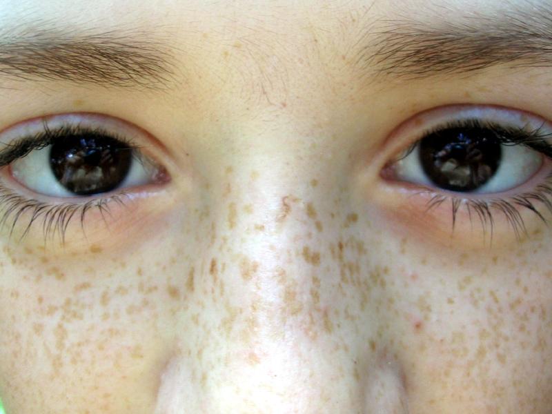
Caption: Freckles are small, flat spots that often become darker with ultraviolet exposure and lighter when exposure reduces.
Image Source: WikiDoc
How do freckles happen?
Freckles are like tiny sun-sensitive paint dots. The number of melanocytes is not greatly increased. Instead, the melanocytes already present make more melanin when the skin is exposed to ultraviolet light.
Genetics matters. Freckles commonly begin in childhood and are more noticeable in people with fair skin, although they can occur across skin tones.
What do they look like?
- Small, flat tan or light-brown spots
- Usually evenly coloured
- Common across the nose, cheeks, shoulders, chest and arms
- Often darker or more numerous in summer
- May fade during winter or with careful sun protection
- Usually present as many similar-looking spots rather than one unusual spot
Freckles do not usually cause pain, itching or scaling.
How are freckles treated?
Freckles are harmless and do not need treatment. Daily broad-spectrum SPF 50+ sunscreen, shade, hats and protective clothing may help prevent them from becoming darker and reduce the development of additional sun-related pigment.
If a patient wishes to soften their appearance, camouflage, selected topical ingredients, intense pulsed light or pigment-targeting laser may be considered after assessment. Results vary and freckles may return with further light exposure. Treatment should never be used as a substitute for appropriate skin checks.
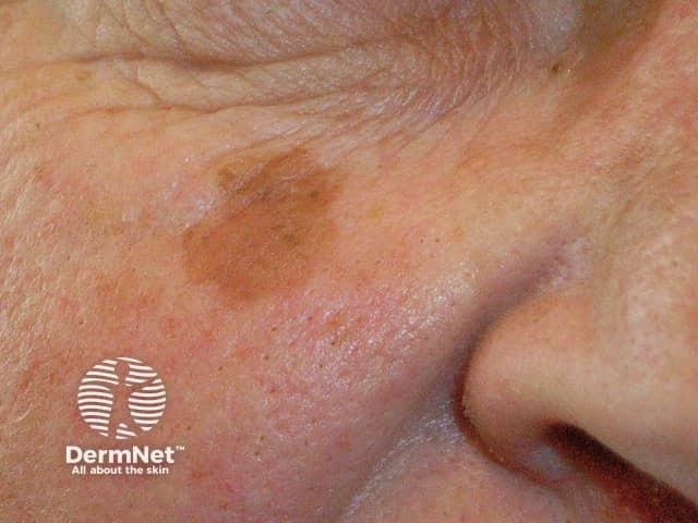
2. Sun spots, or solar lentigines
Caption: Solar lentigines are persistent, well-defined areas of pigment that develop after cumulative ultraviolet exposure.
Image Source: DermNet Lentigo images
How do sun spots happen?
Sun spots are a record of accumulated light exposure. Imagine that one small patch of skin has been repeatedly asked to make extra umbrellas for many years. Eventually, that patch develops a lasting increase in pigment and changes in the way the surface skin cells grow.
Unlike freckles, sun spots usually remain visible throughout the year.
What do they look like?
- Flat, round, oval or gently irregular spots
- Usually tan, brown or dark brown
- More clearly defined than melasma
- Often larger than freckles
- Common on the face, backs of the hands, forearms, shoulders and upper chest
- Usually persist through winter
- Become more common with age and cumulative sun exposure
Solar lentigines are usually harmless. However, an irregular lentigo can resemble melanoma or another sun-related lesion. A spot that is new, changing, unusually dark, multicoloured or different from the others requires medical assessment.
How are sun spots treated?
Treatment begins with confirming the diagnosis. Broad-spectrum SPF 50+ sunscreen and physical sun protection may help prevent new lesions and may modestly reduce further darkening.
Depending on the patient’s skin, the depth and pattern of pigment, options may include topical pigment-modulating care, chemical peels, cryotherapy, intense pulsed light or pigment-targeting laser. A treatment suitable for one isolated sun spot may be unsuitable for melasma or an undiagnosed lesion. Recurrence and new spots remain possible because treatment does not erase the skin’s history of ultraviolet exposure.
3. Melasma
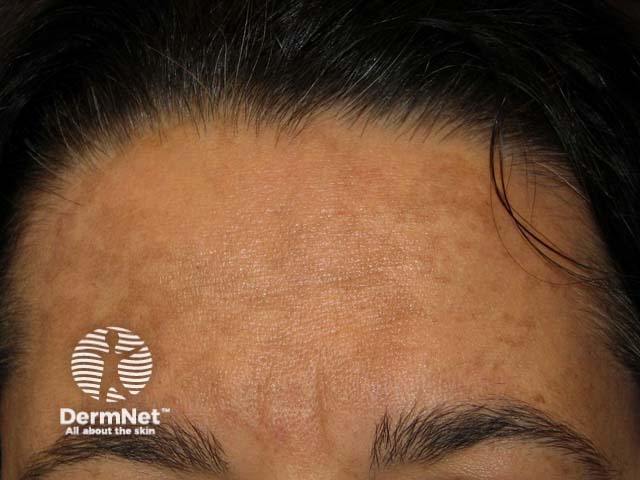
Caption: Melasma usually appears as symmetrical, patchy brown or grey-brown pigmentation on sun-exposed areas of the face.
Image Source: DermNet Melasma (facial pigmentation)
How does melasma happen?
Melasma is not simply pigment sitting on the skin. It is a long-term tendency for the pigment-making system to become overactive.
Imagine that the melanocytes have become very jumpy. Ultraviolet light, visible light, hormones and sometimes heat can ring their alarm bell. Once activated, they may keep making extra pigment even after the trigger has passed.
Pregnancy, some hormonal medicines and a family tendency can contribute. Melasma is more common in women and in skin that tans easily, but it can affect anyone. It is not contagious and it is not caused by poor skin cleansing.
What does it look like?
- Flat, patchy light-brown, dark-brown or grey-brown pigmentation
- Usually appears on both sides of the face in a similar pattern
- Common across the cheeks, forehead, temples and upper lip
- May form larger clouds of colour rather than separate dots
- Often becomes darker after sun exposure
- May worsen during pregnancy or with hormonal change
- Usually has no itch, pain, scale or change in skin texture
Melasma may be epidermal, dermal or mixed. A clinician may use examination, dermoscopy or a Wood’s lamp to help assess its pattern and depth, although no device can predict treatment response perfectly.
How is melasma treated?
Melasma is best thought of as something to control, rather than something that can always be permanently erased.
Management usually has several layers:
- Reduce the alarm signals. Consistent broad-spectrum SPF 50+ sunscreen, hats, shade and sensible heat management are the foundation. Tinted sunscreens containing iron oxides may provide additional protection from visible light for some patients.
- Quiet the pigment-making cells. A doctor may recommend prescription or non-prescription topical ingredients that reduce melanin production or transfer. The choice depends on the patient’s skin, medical history, pregnancy status and tolerance.
- Move existing superficial pigment along. Retinoids, selected acids and other topical agents may help normal skin turnover and gradually improve epidermal pigment.
- Use procedures carefully. Some peels, light-based treatments and lasers may help selected patients, but they can also inflame the skin and worsen melasma or cause post-inflammatory hyperpigmentation. Conservative treatment planning and preparation of the skin are important.
- Plan for maintenance. Even when melasma improves, it can return. A gentle long-term plan is often more useful than repeatedly chasing the pigment with aggressive treatment.
Oral or prescription treatments are not suitable for everyone and require individual medical assessment. Pregnancy and breastfeeding can significantly change which options are appropriate.
4. Post-inflammatory hyperpigmentation
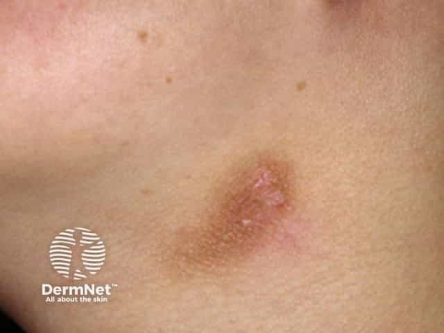
Caption: Post-inflammatory hyperpigmentation follows the exact sites of previous inflammation, such as acne, eczema, injury or irritation.
Image Source: DermNet Postinflammatory hyperpigmentation
How does post-inflammatory hyperpigmentation happen?
Inflammation is like a small fire alarm in the skin. While the skin is repairing itself, that alarm can accidentally tell melanocytes to release extra pigment.
After the pimple, rash, burn or injury has settled, the colour can remain behind like a footprint. If inflammation damages the boundary between the upper and deeper layers of skin, some pigment can drop into the dermis, where it takes longer to clear.
PIH can follow acne, eczema, insect bites, picking, burns, friction, cosmetic procedures or irritating skincare. It can occur in any skin tone, but is often darker and more persistent in skin with a greater natural capacity to make melanin.
What does it look like?
- Flat areas of brown, dark brown, grey-brown or purple-brown colour
- Appears in the same shape and location as the earlier inflammation
- Common after pimples, even when the acne itself has healed
- Does not usually create a new lump or change in skin texture
- Superficial PIH tends to look brown
- Deeper PIH may look grey or blue-grey and fade more slowly
Red or pink marks after acne are called post-inflammatory erythema. They are caused mainly by small blood vessels rather than excess melanin, so they are not treated in exactly the same way.
How is post-inflammatory hyperpigmentation treated?
The first job is to stop new footprints from forming. Acne, eczema, irritation or another underlying inflammatory condition needs to be controlled. Picking, scrubbing and repeatedly irritating the skin can prolong the process.
Daily broad-spectrum SPF 50+ sunscreen helps prevent the marks from becoming darker. Gentle topical ingredients may be used to reduce pigment production and support cell turnover. The right choice depends on the cause of inflammation, pigment depth, skin tone and skin sensitivity.
PIH often fades gradually, but this may take months and deeper pigment can persist longer. Peels, needling, light-based treatment or laser may be considered in selected cases. Poorly chosen or overly aggressive treatment may create more inflammation and therefore more pigment.
5. Deeper dermal pigmentation
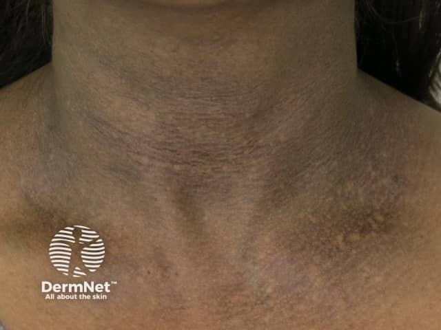
Suggested caption: Blue-grey colour can suggest that pigment sits more deeply in the dermis, but diagnosis requires clinical assessment.
Image Source: DermNet Acquired dermal macular hyperpigmentation
How does deeper pigment happen?
When brown melanin sits deeper in the skin, light travels through more tissue before it returns to the eye. This can make the pigment look grey, slate-coloured or blue-grey.
Examples include dermal melasma, deeper PIH and dermal melanocytosis such as acquired bilateral naevus of Ota-like macules. These conditions have different causes and should not be grouped together simply because they appear grey.
What does it look like?
- Grey-brown, slate-grey or blue-grey colour
- May appear as small speckles or larger patches
- Often changes less from season to season than freckles
- May respond poorly to creams designed for superficial pigment
- Requires careful distinction from melasma, PIH, medication-related pigmentation and melanocytic lesions
How is deeper pigment treated?
Treatment depends entirely on the diagnosis. Topical care may have limited effect when pigment is deep. Selected pigment-targeting lasers may be considered for particular conditions, usually as a course of conservative treatments. Improvement may be gradual and incomplete, and there is a risk of inflammation, lightening or darkening of the skin.
When a brown spot may not be “just pigment”
Not every brown mark belongs in a cosmetic treatment plan. Moles, seborrhoeic keratoses, pigmented actinic keratoses and skin cancers can all be mistaken for pigmentation.
Arrange medical assessment if a spot is:
- New or changing
- Different from the patient’s other spots, sometimes called the “ugly duckling” sign
- Asymmetrical
- Irregular at the edge
- Made of several colours
- Becoming larger, thicker or raised
- Persistently rough, scaly or crusted
- Itchy, painful, bleeding, oozing or not healing
A cosmetic consultation does not replace a comprehensive skin cancer check. If the diagnosis is uncertain, treatment should pause until the lesion has been appropriately assessed.
Why the wrong treatment can make pigment worse
Pigment does not always need stronger treatment. Often, it needs more accurate treatment.
Heat, irritation and inflammation can activate melanocytes. An aggressive peel, laser or home skincare routine may remove some surface pigment while creating a new inflammatory signal underneath. This is particularly important in melasma, sensitive skin and skin tones that develop PIH easily.
A thoughtful plan asks:
- What is the diagnosis?
- Is the pigment superficial, deep or mixed?
- Is the pigment-making cell still being stimulated?
- Is there active acne, dermatitis or irritation?
- How does this patient’s skin respond to inflammation?
- Are pregnancy, hormones, medicines or medical conditions relevant?
- Is the treatment likely to help more than it harms?
Sometimes the safest first treatment is not a procedure. It may be controlling inflammation, simplifying skincare and protecting the skin from light while its behaviour is observed.
The treatment ladder
Pigmentation treatment usually works best from the bottom of the ladder upwards.
Step 1: Protect
Broad-spectrum SPF 50+ sunscreen, hats, shade and protective clothing reduce ultraviolet exposure. For melasma and some forms of hyperpigmentation, a suitably tinted sunscreen may also help protect against visible light.
Step 2: Settle
Active acne, eczema, irritation and an impaired skin barrier need attention. A calm skin barrier is less likely to generate new inflammatory pigment.
Step 3: Regulate
Selected topical ingredients may reduce melanin production, reduce pigment transfer or improve skin-cell turnover. These may include prescription and non-prescription options. More ingredients are not always better, especially if the combination causes irritation.
Step 4: Treat selectively
Peels, intense pulsed light, resurfacing treatments and pigment-targeting lasers can have a role after diagnosis and skin preparation. The most appropriate device and settings depend on the pigment type, depth, skin tone and risk of PIH.
Step 5: Maintain
Pigment can return because melanocytes remain part of the skin. Maintenance usually includes light protection, a tolerable skincare plan and early review if pigment begins to reactivate.
The most important lesson
The word “pigmentation” describes a colour, not a diagnosis.
Freckles, sun spots, melasma and post-inflammatory marks can all be brown, but the reason for the colour is different. When the cause is understood, treatment can be calmer, more precise and less likely to create another problem.
Atelier’s approach begins with assessment and education. The aim is not to promise perfectly even, pigment-free skin. It is to help each patient understand what their skin is doing, which options may be appropriate, and when leaving the skin alone is the better decision.
Not sure which type of pigmentation you have?
A consultation can help identify the likely pattern of pigmentation, assess factors that may be contributing to it, and explain which treatment options may be suitable for your skin. If a lesion requires a dedicated skin cancer assessment, this will be discussed before cosmetic treatment is considered.
References
- Australasian College of Dermatologists. Melasma. https://www.dermcoll.edu.au/atoz/melasma/
- Doolan BJ, Gupta M. Melasma. Australian Journal of General Practice. 2021;50(12). https://www1.racgp.org.au/ajgp/2021/december/melasma
- DermNet. Melasma. https://dermnetnz.org/topics/melasma
- DermNet. Ephelis. https://dermnetnz.org/topics/ephelis
- DermNet. Solar lentigo. https://dermnetnz.org/topics/solar-lentigo
- DermNet. Postinflammatory hyperpigmentation. https://dermnetnz.org/topics/postinflammatory-hyperpigmentation
- Mar K, et al. Treatment of post-inflammatory hyperpigmentation in skin of colour: a systematic review. 2024. https://pmc.ncbi.nlm.nih.gov/articles/PMC11514325/
- Sarkar R, et al. Delphi consensus on melasma management by international experts and pigmentary disorders society. Journal of the European Academy of Dermatology and Venereology. 2025/2026. https://pubmed.ncbi.nlm.nih.gov/40996222/
- Cancer Council Australia. Check for signs of skin cancer. https://www.cancer.org.au/cancer-information/causes-and-prevention/sun-safety/check-for-signs-of-skin-cancer
- Cancer Australia. What are the symptoms of melanoma? https://www.canceraustralia.gov.au/cancer-types/melanoma-skin/what-are-symptoms-melanoma
Medical disclaimer
This information is provided for general educational purposes only. It does not constitute personal medical advice and should not replace an individual, in-person consultation with an appropriately qualified healthcare practitioner. Treatment suitability, expected outcomes and potential risks vary between patients and require assessment of your medical history, clinical findings and individual circumstances.